Male Reproductive System Histology
Male Reproductive System Lab
Learning Objectives
- Describe the histological organization of the testis and the process of spermatogenesis in the germinal epithelium of the seminiferous tubule
- Contrast spermatogenesis from spermiogenesis
- Draw a sperm cell and label its major parts
- Explain the importance of each portion of the duct system and accessory glands of the male reproductive tract
- Explain the structural and functional significance of the blood-testis barrier
- Describe the structure and function of the prostate gland
Pre-Lab Reading
Introduction
The male reproductive system can be thought of as a series of tubes. These tubes deliver the male gametes from their site of production in the testes to their destination outside the body. The system itself is divided into two distinct units: testes, located outside the major body cavity and housed in the scrotum and the excretory duct system, which transports the sperm from the testes and whose accessory glands produce and modify the contents of semen.
Testes
The testes are a source of gametes and steroid sex hormones. It is encapsulated by the fibrous tunica albuginea and tunica vasculosa (not visible here). Septa extending inwards from the tunica albuginea partition the gland into lobules. The bulk of the gland is composed of the seminiferous tubules, in which sperm develop. After exiting the testicular duct system composed of the rete testis and ductus efferentes, spermatozoa enter the highly convoluted epididymis, visible on the dorsal aspect of the testis here. 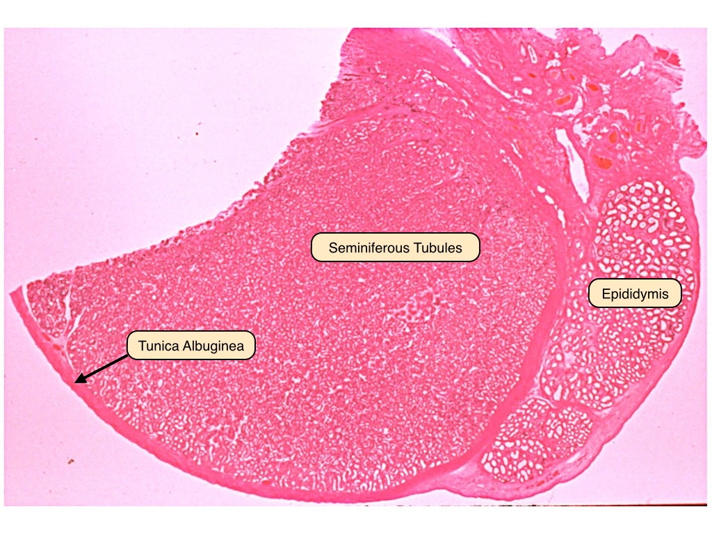
Seminiferous Tubules
Spermatozoa are produced in the germinal epithelium of the seminiferous tubules and released into the lumina of these ducts. The germinal epithelium contains both Sertoli cells and the developing spermatocytes. Sertoli cells extend from the basement membrane of the germinal epithelium to the lumen of the tubule. These cells envelope the developing sperm cells. They are joined to one another by junctional complexes and form the blood-testis barrier. The interstitial space contains clumps of darker, eosinophilic cells. These are the Leydig cells, which produce and release testosterone. Myoid cells surround the tubules and generate rhythmic contractions to propel spermatozoa and fluid. They also synthesize collagen and other fibers of connective tissue. 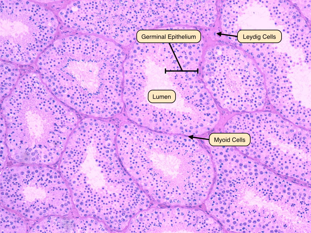
Spermatogenesis
This is magnified image of the germinal epithelium. The epithelium rests on a basement membrane and surrounds a lumen where sprematozoa are released. Identify the spermatogonia, located in the basal compartments of both membranes. These cells appear round and pale, with prominent nucleoli. Sertoli cells, with their characteristic oval-shaped nuclei, are also visible. These provide support to the developing primary spermatocytes, which have large, granulated nuclei that are preparing for the first meiotic division. Secondary spermatocytes, which contain 23 pairs of chromatids, are rarely visible. The products of meiosis are the haploid spermatids, which contain dark, round nuclei and a decreasing amount of cytoplasm. These differentiate further into spermatozoa. Remember that cytokinesis is incomplete during these steps, and cytoplasmic bridges connect the cells and allow for their synchronous development. 
Leydig Cells
Interstitial or Leydig cells are located in the connective tissue surrounding the seminiferous tubules. They produce testosterone, the male sex hormone responsible for the growth and maintenance of the cells of the germinal epithelium and the development of secondary sex characteristics. Leydig cells often display cytoplasmic crystals of Reinke, the function of these crystals is unknown. 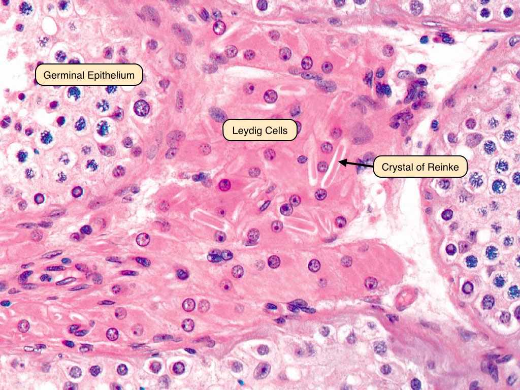
Sertoli Cells
Sertoli cells are located in the germinal epithelium and play a supportive role in the development of spermatozoa. These cells have abundant cytoplasm and extend from the basement membrane to the lumen. Sertoli cells have a characteristic oval nucleus with a dark nucleolus. The cytoplasmic contents and blood-testis barrier are better visualized under the electron microscope. 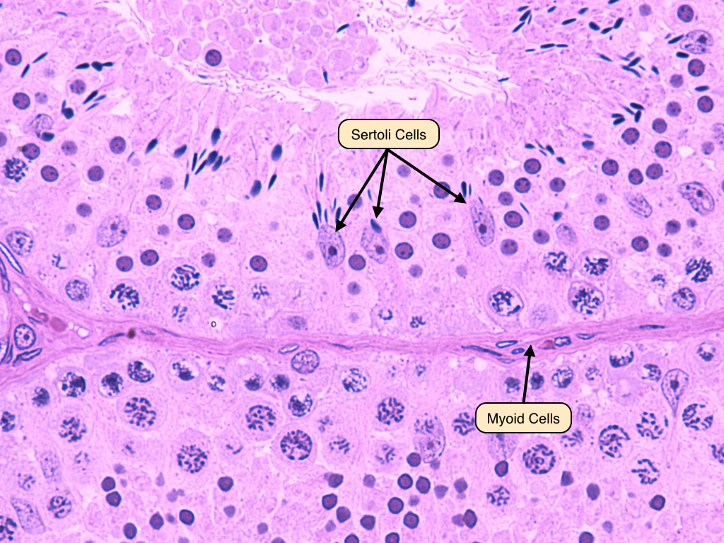
Rete Testis
The rete testis connects the seminiferous tubules to the ductus efferentes. It is lined by ciliated cuboidal epithelial cells that also contain microvilli. The activity of the cilia helps to move the spermatozoa along the tube, as they are immobile until they reach the epididymis. The microvilli absorb excess materials, including protein and potassium, from the seminal fluid. 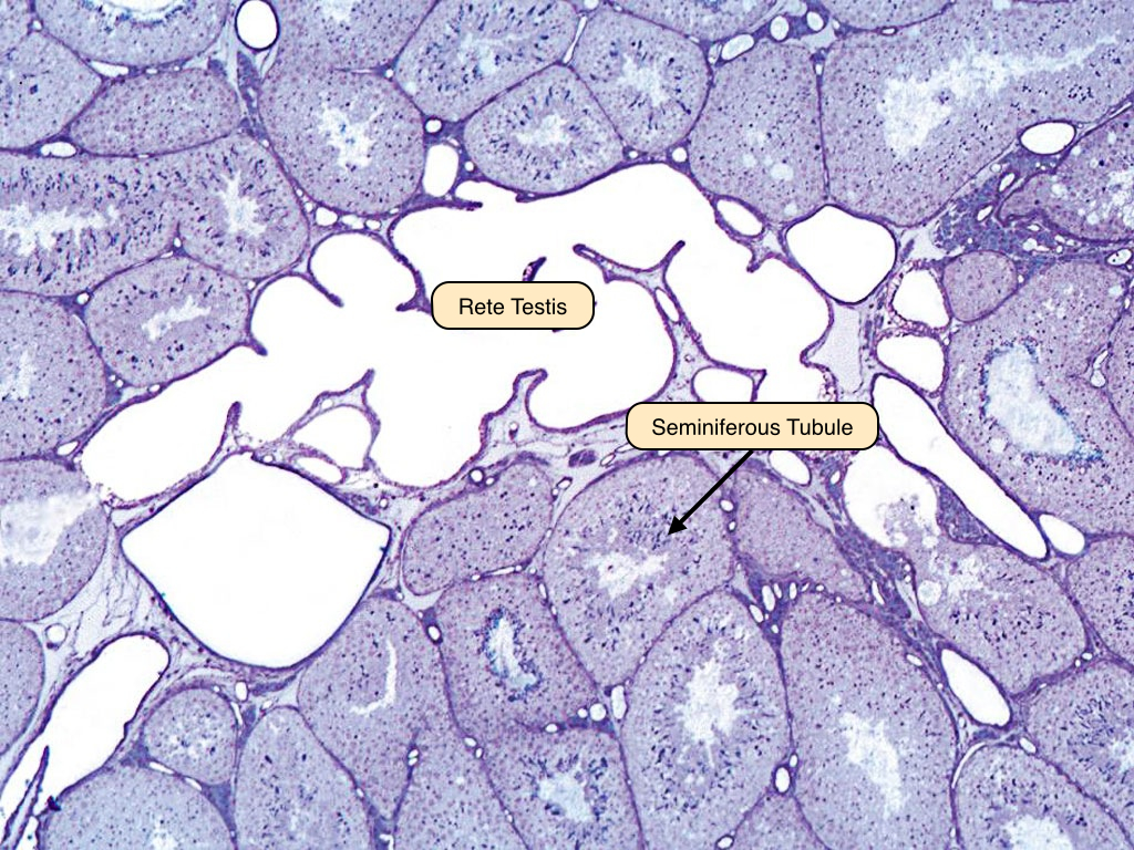
Ductuli Efferentes
The ductuli efferentes emerge from the dorso-superior margin of each testis. They originate from the rete testis and gradually fuse to form the ductus epididymis. The epithelium has a characteristic scalloped appearance that results from a lining that contains both cuboidal and columnar epithelial cells. A layer of smooth muscle surrounds the walls. The non-ciliated cells reabsorb testicular fluid, while the ciliated cells propel the immobile sperm to the epididymis, where they gain the ability to swim. 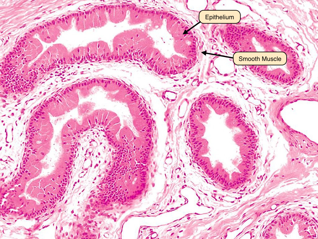
Epididymis
Each epididymis is formed by a single convoluted tubule seen in multiple cross sections. It is lined by a tall pseudostratified columnar epithelium. These cells bear stereocilia on their luminal surface that absorb fluid released from the testes along with sperm. In this section, the spermatozoa can be seen in the lumen throughout the epididymis. 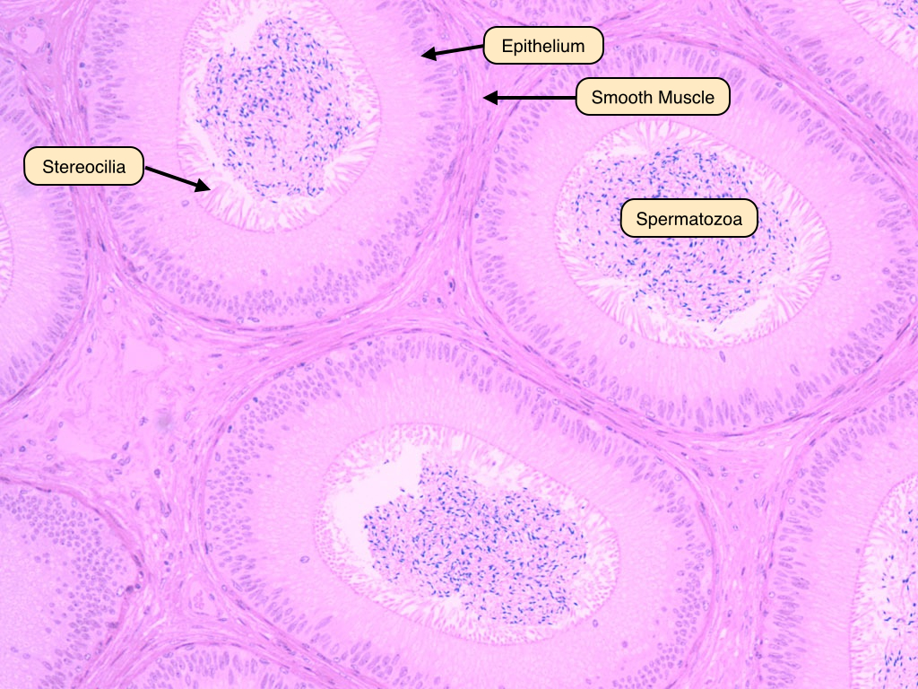
Ductus Deferens
The ductus deferens extends from the epididymis to the ejaculatory ducts. The epithelium of this tube displays a pseudostratified columnar epithelium and is surrounded by a prominent muscular layer. This layer contains inner and outer longitudinal muscle and middle circular muscle. An adventitia of connective tissue surrounds the muscularis layer. 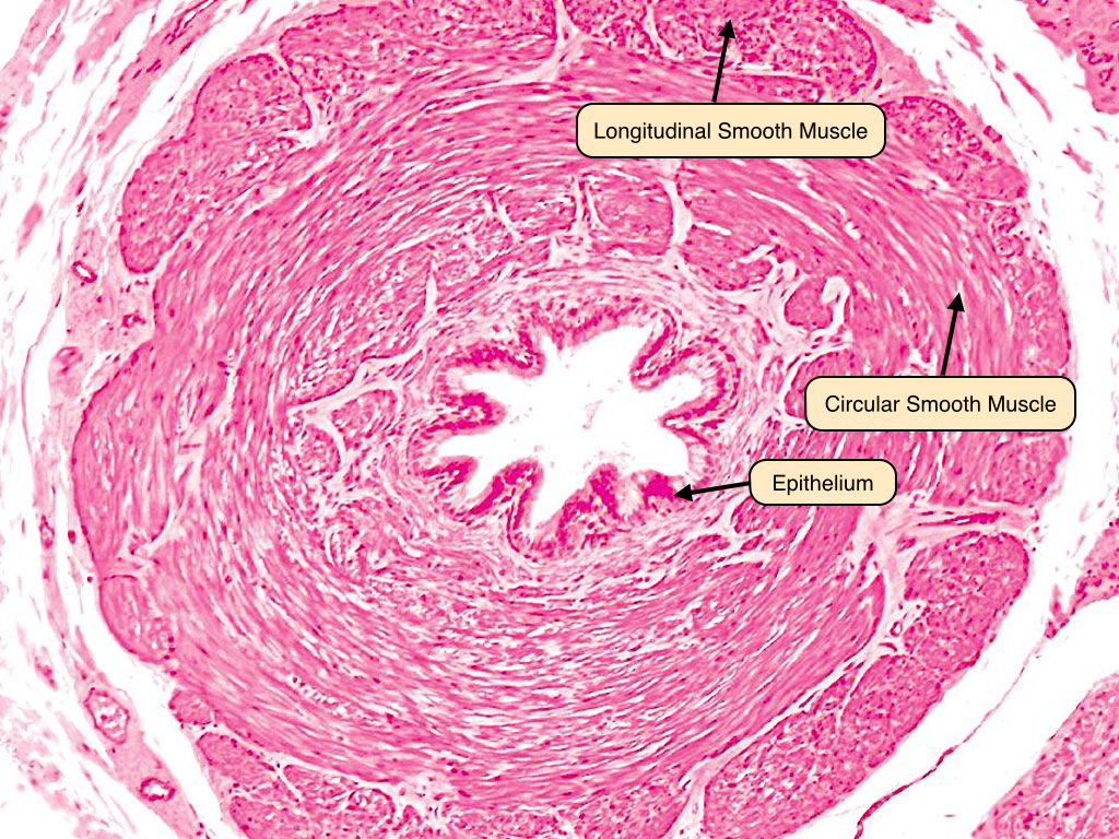
Urethra
The human urethra is lined by a pseudostratified columnar epithelium lining the urethral lumen. The urethra is located in the corpus spongiosum made up of erectile tissue. Note the blood vessels contained in the erectile tissue; during erection, the arteries dilate to fill the sinuses, which obstruct venous outflow and traps blood in the penis. 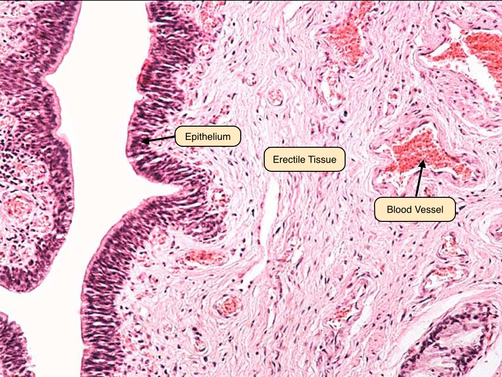
Seminal Vesicle
The seminal vesicles appear as honeycombed saccules with thin, highly branched folds of mucosa, lined by a pseudostratified columnar epithelium. These folds join one another to delimit irregular spaces, which communicate with a large, central lumen filled with a pale-staining, homogenous secretion. Observe the coat of smooth muscle surrounding the saccular dilation of the gland. Its contraction expels the accumulated secretion during ejaculation. 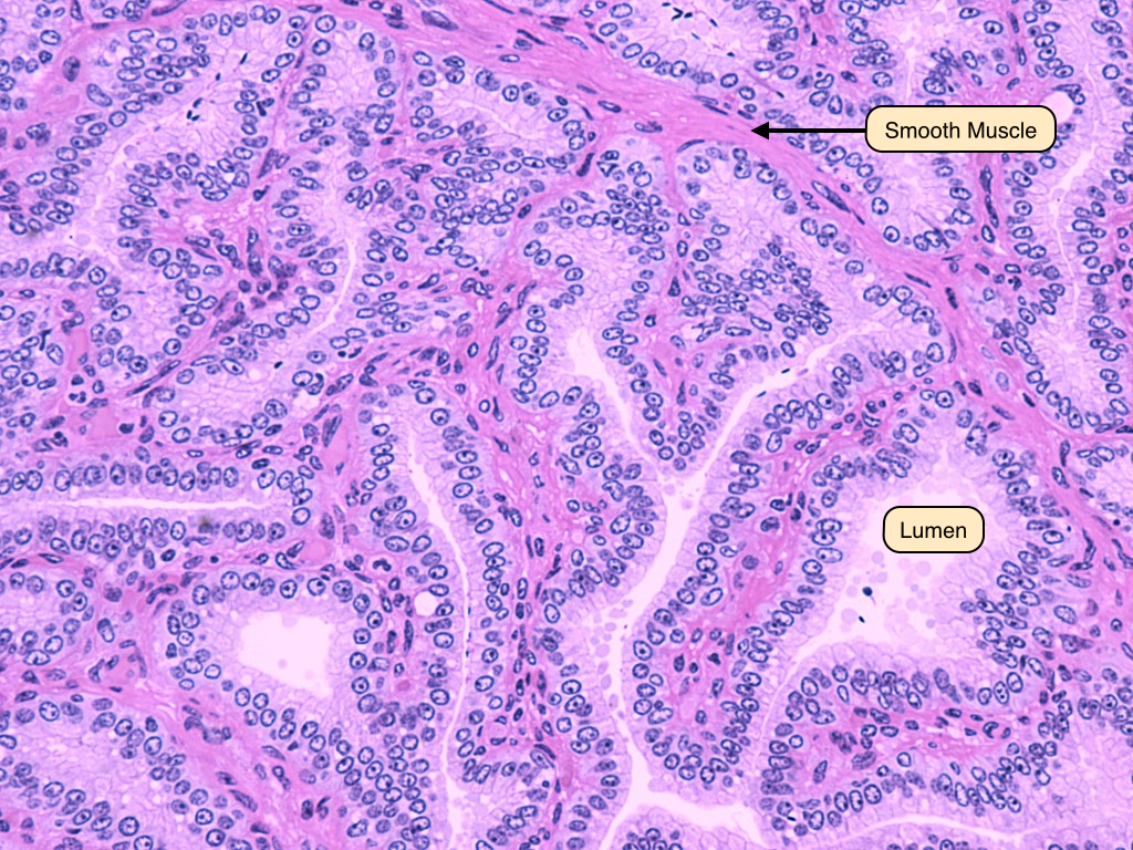
Prostate Gland
The prostate is a walnut-sized conglomeration of tubulo-acinar glands that surrounds the initial segment of the urethra. The epithelium of the prostate produces a fluid rich in citric acid and proteolytic enzymes that nourish and prevent the coagulation of sperm in the vagina. Characteristic of the gland are prostatic concretions. These accumulate over time in the lumina of the tubulo-alveolar glands. Note the presence of numerous basal cells in the epithelium of the glands. Their presence distinguishes benign and malignant glands. 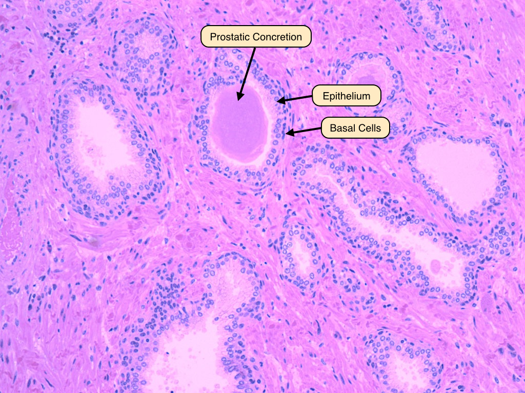
Penis
The penis contains three columns of erectile tissue: two corpus cavernosa and a single corpus spongiosum containing the penile urethra. The erectile tissue of the penis appears as a vast sponge-like system of irregular vascular spaces intercalated between the arteries and veins. These sinuses receive blood from the helicine arteries, which dilate during erection to engorge the sinuses with blood. This, in turn, restricts venous outflow.Note the vast sponge-like arrangement of irregular vascular spaces intercalated between the arteries and veins. 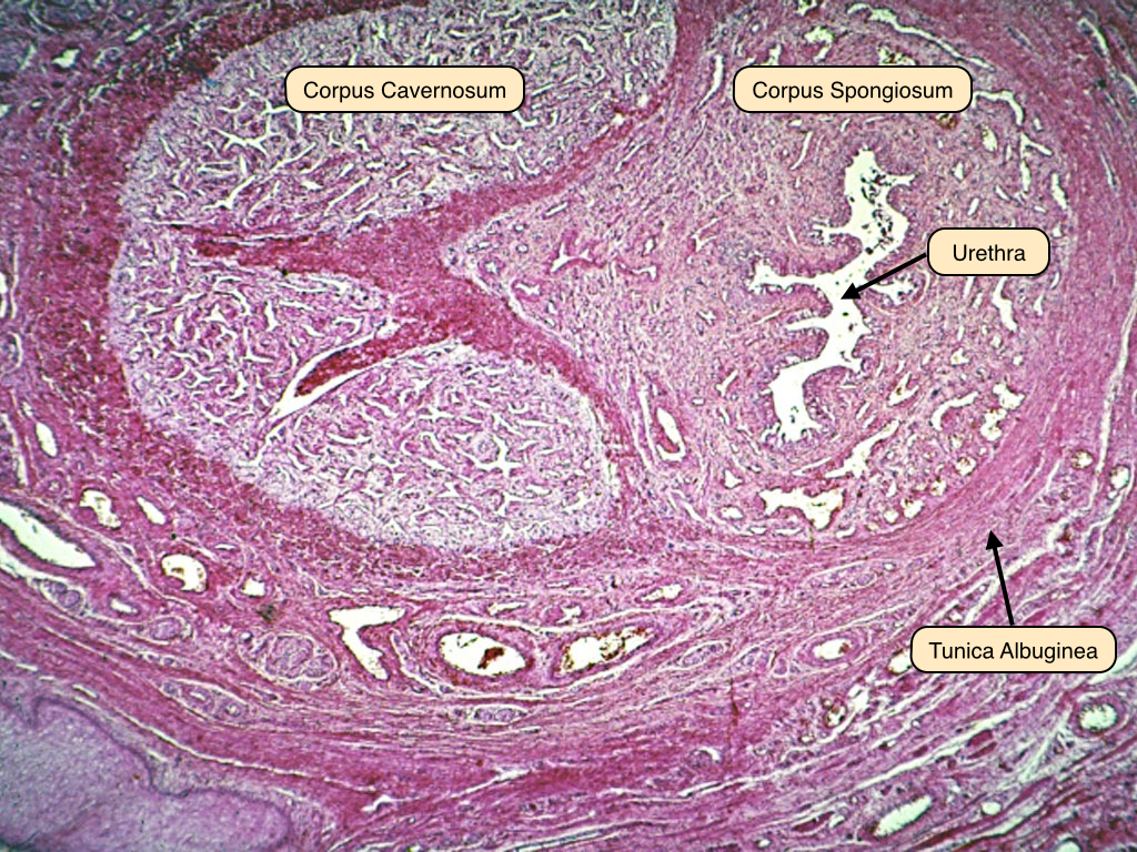


No comments:
Post a Comment