Monday, February 26, 2024

application 11

Scientist Spotlight: Augustus White


Augustus White's biography includes, among other things, attending segregated schools as a boy, playing football in college, and receiving the Bronze Star for his service in the Army Medical Corp in Vietnam. He was the first African American to graduate from Stanford's school of medicine and the first African American resident in orthopedics at Yale. His research focuses on the biomechanics of the bones - especially the spine - and fracture healing. Dr. White additionally leads efforts to promote diversity in science and to explore the roles of unconscious bias in medicine.


1) Please click here Links to an external site. to read an article on Dr. White's biography. 


2) Now review the following two videos on the general topics of Dr. White's work: bone remodeling and fracture repair. 

Click hereLinks to an external site. (or copy and paste this address https://www.youtube.com/watch?v=Ux7xsXihQnY) to watch the video regarding bone remodeling. Then click hereLinks to an external site. (or copy and paste this address https://www.youtube.com/watch?v=-P6LsendHxU) to view an animation of bone fracture repair (set to some very dramatic piano music!). Note in particular what types of cells and molecules are involved and what those cells seem to be doing at different stages in these processes. Do not worry about some of the less-familiar molecule names, like all the growth factors mentioned.


After reviewing these resources, write a 250 word or more reflection with your responses. You might wish to discuss any or all of the following topics:


1)   What was most interesting or most confusing about the these resources?


2)   What can you learn from these resources about the physiology of bone remodeling and fracture healing?


3)   What do these resources tell you about the types of people that do science?


4)   What new questions do you have after reviewing these resources?

Week 7 - Estrogen & Bone: Elaborate/Evaluate

  As a result of menopause, which of the following imbalances is likely to occur?

  1.   osteocytes will begin dividing frequently
  2.   osteoblast activity will outpace osteoclast activity
  3.   osteoclast activity will outpace osteoblast activity
  4.   osteoblasts will begin dividing frequently

 Though this treatment comes with a number of potentially serious side effects, one popular treatment for osteoporosis has been hormone replacement therapy.  In that therapy, the patient takes medications containing estrogen.  The increase in estrogen levels would help treat osteoporosis by...

  1.   Increasing the lifespan of osteoclasts
  2.   Increasing the lifespan of osteoblasts
  3.   Encouraging cell division in osteoclasts
  4.   Causing apoptosis in osteblasts


Week 7 - Physical Activity Feedback Loop: Explain

 Background Fact: Older individuals are sometimes less physically active than younger individuals, which makes older individuals more at risk for osteoporosis. Interestingly, this is the same phenomenon that makes astronauts at risk for osteoporosis if spending long periods in space.
Brainteaser: Try to reason out a feedback loop that could explain how a decrease in physical activity could worsen osteoporosis.

Perhaps the main thing to consider here is how the bones know that they are experiencing less physical activity and should therefore change their strength.  In other words, what is the stimulus and the receptor in this feedback loop?  It turns out, this comes down to one of our main bone cells: the osteocyte!  Below is a brief excerpt from a scientific article published in 2012, titled  Osteocyte Signaling in Bone

 Osteocytes, the cells residing within the bone matrix and comprising 90% to 95% of the all bone cells, have long been considered quiescent bystander cells compared to the osteoblasts and osteoclasts whose activities cause bone gain and loss, and whose dysfunction lead to growth defects and osteoporosis. However, recent studies show that osteocytes play a crucial, central role in regulating the dynamic nature of bone in all its diverse functions. Osteocytes are now known to be the principal sensors for mechanical loading of bone. They produce chemicals that regulate bone formation and resorption. Osteocytes appear to be the major local orchestrator of many of bone’s functions."

Note in particular that osteocytes sense "mechanical loading," or the amount of pressure on our bones.  This sort of makes sense if you look at the picture of osteocytes above.  Those cells are squeezed in small gaps within compact bone.  You could imagine, then, that pressure placed on the bones would cause osteocytes to experience more "squeezing" in their small spaces.

lifting weightsAs the quote above points out, the osteocytes are then able to signal osteoclasts and osteoblasts to adjust bone thickness appropriately.  If you start lifting weights and putting extra pressure on your bones, your osteocytes will sense that and tell your osteoblasts to add more bone to make the bones stronger.  With stronger bones, the pressure would then be relieved.

reduced physical activityOn the other hand, if you stop performing as much physical activity (or go into orbit around Earth where you experience less gravity!), your bones would experience less pressure.  Your osteocytes would sense that and signal your osteoclasts to tell them less bone is required to meet the body's needs.  Your osteoclasts might then remove bone, which would create weaker bones and bring the pressure on the bones back up to normal levels.  Of course, that could also contribute to osteoporosis!

 Background Fact: Older individuals are sometimes less physically active than younger individuals, which makes older individuals more at risk for osteoporosis. Interestingly, this is the same phenomenon that makes astronauts at risk for osteoporosis if spending long periods in space. Space Shuttle Endeavour on Pad39A

Brainteaser: Try to reason out a feedback loop that could explain how a decrease in physical activity could worsen osteoporosis.

Take some time to consider the above scenario.  Think in particular about what cell types might be responsible for creating the condition of osteoporosis in this situation.  You're welcome to look for some answers online to explain this, but don't spend more than a few minutes doing so!

Once you have taken some time to think and find information, submit a few sentences here to record your thoughts.  You might discuss your ideas about what interactions among cells serve to create weak bones after a lack of physical activity, or just talk about what you find confusing/questions you have!

  Score for this attempt: 5 out of 5
Submitted Feb 26 at 4:05pm
This attempt took less than 1 minute.
 
Question 1
1 / 1 pts
Which of the following organs require calcium in order to function appropriately? (check all that apply)
  heart
  brain
  muscle
  bones
 

Question 2
1 / 1 pts
Which of the following has the receptors that detect blood calcium levels?
  parathyroid gland
  osteoclast
  osteoblast
  PTH
  bone
 
Question 3
1 / 1 pts
Which of the following is the most direct representation of the efferent pathway in the feedback loop that occurs in response to low blood calcium?
  osteoclast
  osteoblast
  parathyroid gland
  PTH
  raised blood calcium levels
 
Question 4
1 / 1 pts
Given everything we've studied so far in this module, what is the most likely explanation for how a person could have osteoporosis at the same time as having normal blood calcium?
  the body is taking calcium from bones in order to keep blood calcium normal
  the osteoblasts have lost their ability to effectively incorporate calcium into bone
  the parathyroid gland is interfering with Vitamin D's ability to assist in calcium absorption
  the digestive system has become insensitive to Vitamin D
  the osteoclasts are malfunctioning and breaking down bone tissue inappropriately
 
Question 5
1 / 1 pts
The feedback loop we just studied that (in our scenario) resulted in osteoporosis is an example of a...
  Rheostasis
  Positive Feedback Loop
  Negative Feedback Loop


Deviation From  Normal : Low level of Vitamin D

Receptor                         : Bone Cell

Control Center               :parathyroid gland

Efferent Pathway           :parathyroid hormone (effects on bone because of inhibition production of hormone)

Effector                          :Stimulating Osteoclasts and inhibiting  Osteoblasts

Response                       : When blood calcium levels are low, parathyroid hormone (PTH)is released, PTH stimulates osteoclasts , also PTH inhibits  osteoblasts.Osteoclasts break down bone tissue, releasing calcium into the bloodstream.PTH also inhibits osteoblasts, which are cells involved in bone deposition. By reducing calcium deposition in bone leading not enough calcium, our bones become more fragile, leading to osteoporosis.

Sunday, February 25, 2024

Week 7 - Vitamin D Feedback Loop: Explain 2

 Week 7 - Vitamin D Feedback Loop: Explain 2

As a reminder, we are discussing the following brainteaser:

osteoporosisBackground Fact: Vitamin D is important for the absorption of calcium from food.
Brainteaser: Try to reason out how low Vitamin D could worsen osteoporosis in an individual, even if that person has normal blood calcium levels.
Feedback Loop:

Picking up from the last page, we're trying to figure out how the body would find extra calcium if blood calcium levels drop, and also how that would relate to osteoporosis.

View the first few minutes of the following animation to learn about the feedback loop that begins when blood calcium levels fall (or copy and paste this address: https://www.youtube.com/watch?v=tQVzZaV9yDY)

Links to an external site.  Note that you only need to review and fully understand the portion from the start up until 2:15.

Now review the video a second time (again, just until 2:15) and try to figure out the components of the feedback loop.  That is, try to identify which cells, organs, or molecules are serving as receptors, control center, efferent pathway, and effectors.  Take out a scratch paper and try matching each role to a body part/cell/molecule!  Also consider what would be the deviation from normal and the response.  You can also refer to the bottom half of the textbook diagram below.

In this illustration, the two mechanisms that maintain calcium homeostasis are shown as two semicircles that are combined, one on top of each other, to make a complete circle. Homeostasis occurs along the diameter of the circle, at the border between the two semicircles. At homoeostasis, normal calcium levels are 10 milligrams per deciliter. The upper semicircle represents the mechanism that reduces elevated calcium levels in the blood when the levels are too high. First, the thyroid gland releases calcitonin. Calcitonin activity inhibits osteoclasts and decreases the reabsorption of calcium ions in the kidney. These two actions cause calcium ion levels in the blood to drop back to homeostasis. The lower semicircle represents the mechanisms that increase calcium levels in the blood when the levels are too low. First, the parathyroid glands release PTH. PTH stimulates osteoclast activity, causing calcium ions to be released from bone. PTH also increases the reabsorption of calcium by the kidney. In addition, PTH also increases calcium absorption in the small intestines via Vitamin D synthesis. These three actions cause calcium ion levels in the blood to increase.

Scientist Spotlight #6

 We will next move on to bone physiology and will begin our studies by getting to know the work of Mercedes Lopez, a Principal Investigator at the National Polytechnic Institute in Guanajuato, Mexico.  Her research focuses on how naturally occurring chemicals from agave plants (seen in picture with Dr. Lopez) impact human health.  Agave is probably most famously known as the type of plant used in the production of tequila and agave nectar is often used as a sweetener.  In 2014, Dr. Lopez coauthored a study titled Agave Fructans: Their Effect on Mineral Absorption and Bone Mineral Content.  In the summer of 2017, this article was covered by a science news website and the story was subsequently picked up by numerous newspapers and television stations across the country.

1) Please click here
Download click here to read a summary of Dr. Lopez's fascinating path to her current position, which begins... “Mercedes, you cannot learn English and Science at the same time.”  “Guess what? My family had very little, I was the first in my family to ever study outside of Mexico, I am dyslexic, and I DID IT.”

2) Then, click here Links to an external site. to read the science news article that set off a storm of publicity around Dr. Lopez's work.

3) Finally, please briefly review the abstract of the research article

Links to an external site..

After reviewing these resources, write a 250 word or more reflection with your responses. You might wish to discuss any or all of the following topics:

1)   What was most interesting or most confusing about the these resources?

2)   What can you learn from these resources about bone homeostasis and the chemicals important to bone physiology?

3)   What do these resources tell you about the types of people that do science?

4)   What new questions do you have after reviewing these resources?

Hematopoiesis

 Hematopoiesis – the formation of blood cellular components – occurs during embryonic development and throughout adulthood to produce and replenish the blood system. Studying hematopoiesis can help scientists and clinicians to understand better the processes behind blood disorders and cancers.

application 9

From an anatomy and physiology perspective collagen, elastin, keratinocytes, and lipids significantly influences how our skin ages.

(1)Aging turn out the skin loses elasticity because the production of collagen and elastin decreases. Wrinkles are folds or wrinkles that develop on the face and body also presenting significant symptoms such as thinning , sagging skin, wrinkles and dry skin. 

(2)Age-related changes affecting keratinocytes which played vital role for skin strength and flexibility as we age reduction of keratin produce caused thinner epidermis and weaken the skin.

(3)The skin loses its youthful elasticity,wrinkles are folds or wrinkles that develop on the face and body. due to reduced production of two essential proteins: collagen and elastin.

(4)Lipids, including fats and oils, are essential for maintaining skin hydration and barrier function. As we age, lipid production diminishes, resulting in dryness, roughness, and compromised skin barrier integrity. 

Although we cannot stop the natural aging process, according to the article we can adopt preventive measures that could help improve skin health and maintain moisture  by using anti-aging skin cream , serum , moisturizer. Also we should eat well balance nutrition, ensure enough hydration, apply moisturizer, sun protection,   manage stress reduction, reduce smoking, perform regular exercise , get enough sleep to maintain healthy skin and combat the effects of aging.

Thursday, February 22, 2024

Vitiligo

 Vitiligo Individuals with vitiligo experience depigmentation that results in lighter colored patches of skin. The condition is especially noticeable on darker skin. (credit: (a) Klaus D. Peter (b) Owl Bridge Media / Wikimedia.)

melanin

 Albinism is a genetic disorder that affects (completely or partially) the coloring of skin, hair, and eyes. The defect is primarily due to the inability of melanocytes to produce melanin. 

Individuals with albinism tend to appear white or very pale due to the lack of melanin in their skin and hair. Recall that melanin helps protect the skin from the harmful effects of UV radiation. Individuals with albinism tend to need more protection from UV radiation, as they are more prone to sunburns and skin cancer. They also tend to be more sensitive to light and have vision problems due to the lack of pigmentation on the retinal wall. Treatment of this disorder usually involves addressing the symptoms, such as limiting UV light exposure to the skin and eyes. In vitiligo, the melanocytes in certain areas lose their ability to produce melanin, possibly due to an autoimmune reaction. This leads to a loss of color in patches (Figure 5.10). Neither albinism nor vitiligo directly affects the lifespan of an individual

fat deposit

 Where the fat is deposited and accumulates within the hypodermis depends on hormones (testosterone, estrogen, insulin, glucagon, leptin, and others), as well as genetic factors.

Cells of the Epidermis

 Figure 5.6 Cells of the Epidermis The cells in the different layers of the epidermis originate from basal cells located in the stratum basale, yet the cells of each layer are distinctively different. EM × 2700. (Micrograph provided by the Regents of University of Michigan Medical School © 2012)

  Epidermis

 The epidermis is epithelium composed of multiple layers of cells. 


The basal layer consists of cuboidal cells, whereas the outer layers are squamous, keratinized cells, so the whole epithelium is often described as being keratinized stratified squamous epithelium. LM × 40. (Micrograph provided by the Regents of University of Michigan Medical School © 2012)

keratinocytes

 The cells in all of the layers except the stratum basale are called keratinocytes. A keratinocyte is a cell that manufactures and stores the protein keratin. Keratin is an intracellular fibrous protein that gives hair, nails, and skin their hardness and water-resistant properties. 

 multiple layer 

connective tissue

deeper layer has vascularized. 

epidermis

dermis irregular connective , sweat gland 

loose connective tissue and fatty tissue

Dermis - four or five layers 

The epidermis is composed of keratinized, stratified squamous epithelium.

deep to superficial 

Skin that has four layers of cells is referred to as “thin skin.” 

From deep to superficial, these layers are the 

  1. stratum basale, 
  2. stratum spinosum,
  3.  stratum granulosum, and 
  4. stratum corneum.        
Most of the skin can be classified as thin skin.

Thick skin” is found only on the palms of the hands and the soles of the feet. It has a fifth layer, called the stratum lucidum, located between the stratum corneum and the stratum granulosum


Tuesday, February 20, 2024

Leukemia

 Leukemia starts in the soft, inner part of the bones (bone marrow), but often moves quickly into the blood. It can then spread to other parts of the body, such as the lymph nodes, spleen, liver, central nervous system and other organs.

Which specimens do you think represent bones in rickets? Why?

Rickets refers to the deficient mineralization alkaline phosphatase (ALP) and defective organization of cartilage at the growth plate and is only seen in children. Because the growth plates at the ends of the long bones are where lengthening of these bones occurs, children with rickets will experience poor height growth.


What is the bone pathology in osteogenesis imperfecta?
Osteogenesis imperfecta (OI) is a genetic disorder of connective tissues caused by an abnormality in the synthesis or processing of type I collagen. It is also called brittle bone disease. It is characterized by an increased susceptibility to bone fractures and decreased bone density.Feb 6, 2023
How does osteogenesis imperfecta affect the bones?
In the most common forms, people with OI have a gene that carries incorrect instructions in one copy of the gene for making collagen, a substance that makes bones strong. The gene causes the body to not make enough collagen or the collagen does not work properly. This leads to weak bones that break easily.

Osteo

 Osteoprogenitor beginning of the bone cell

Created in embryo

Create new bone

Surrounded in bone become osteocytes

Maintain none found in bone tissue diaphysis 

Chondrocytes 

Chond is cartillage 

Chondrocytes build cartilage


Osteoclasts 

WBC fuses osteoclasts many white blood cell together

Last is actually is break down 

Lysosomes break bone down 

Block shape bone

In reality they have indentation 

Need to be the right shape


skin 02-20-2024


Between those two 

Key players that 
Keratin 
Elastin

Lipids are all over the body 
Somehow impacted by aging
A and P perspective bone cells
Condo sites 
Lab bone tissue
Skeletal system 
Osteoporosis clinical or health related cases 
Feedback loop bone physiology 
Compare and contrast 
Related skin pigmentation 
Specifically melanocytes 
Keratinocytes 
Provide pigmentation of the skin
Different of melanin 
Types of melon that produces 
Turn out 
What melon is there for
Folate
Reproduce folate 
Prevent th destruction of folate 
Vit D is  create by UV  
Inhibiting the production of vit D

Melnin absorb UV rays 
Lack of melanin caused->

We want enough pigmentation 
Not too much pigmentation 
Skin cancer 
Protection that we received from melanin
Quote from Jenna Lester
Fair skin highest risk for skin cancer 


Other type preventable 
Genetic differences in that group 

Fair skin
Understanding skin physiology 
Because of the melanin or pigment of thr skin
Fitzpatrick scale
Type 1 to 6
Base on specific skin type Systematically research


Monday, February 19, 2024

 There are three classes of fibrous ECM proteins: collagen, elastin, and fibronectin. Collagen is the most abundant protein in animals. 

Approximately 25% to 30% of human protein mass is from collagen molecules.

Aging issue wrinkles

 Aging Issue: Wrinkles

Skin wrinkling is one of the most visible clinical features in aged skin, which can be exacerbated by exposure to the sun, smoke, or dehydration. The molecular mechanisms underlying skin wrinkling result from a substantial deterioration of the skin matrix molecules, i.e. diminished and disorganized collagen and reduced and distorted elastic fibers 123.

Collagen is the most abundant extracellular matrix (ECM) protein generated by dermal fibroblasts. Type I collagen accounts for about 80% of the dry weight of the dermis and other types (Il, III, IV) are also found in skin tissues.

Our results showed a dose-dependent increase in COL2A1 at the transcriptional level upon methylene blue (MB) treatment (Fig. 6D). MMPs, the

study, we found a significant inhibition of MMP9 expression upon MB treatment (Fig. 6D), suggesting an attenuation of collagen degradation in the ECM. Insulin like growth factor 1 (IGF-1), a hormone secreted by dermal fibroblasts and keratinocytes, upregulates the expression of collagen and inhibits MMP139. The expression of IGF-1 decreases with increasing age10. In this 3D skin model, we have shown an upregulation of IGh-I transcription upon MB treatment (Fig. 6D), which further supports the idea that MB treatment increases collagen in the dermis. Elastin, the second most abundant EC.M component, is a tibrous protein that contributes to 2~4% of the dry weight of the dermis. It provides natural elasticity and strength to human skin and also plays a role in tissue repair341. We found a robust increase in elastin expression by MB, suggesting that MB treatment enhances skin elasticity and improves skin wound healing (Fig. 6A-C). Based on these results, we speculate that by regulating and orchestrating the expression of these ECM genes, MB may reduce the formation of skin wrinkles.


The molecular mechanisms underlying skin wrinkling result from a substantial
deterioration of the skin matrix molecules, i.e. diminished and disorganized
collagen and reduced and distorted elastic fibers.

Collagen is the most abundant extracellular matrix (ECM) protein generatedby dermal fibroblasts.

MB treatment increases collagen in the dermis .

 Elastin, the second most abundant
ECM component, is a fibrous protein that contributes to 2~4% of the dry
weight of the dermis. It provides natural elasticity and strength to human skin
and also plays a role in tissue repair

Sunday, February 18, 2024

Week 6 - Skin Physiology & Aging: Final Evaluate

 According to our article, which of the following is the main change in the skin during aging that causes wrinkle formation?

  Reduced Keratin 

  Reduced Keratin and Lipids 

  Excessive Collagen and Elastin 

  Excessive Keratin and Lipids 

  Reduced Collagen and Elastin 


Question 2

1 / 1 pts

According to our article, which of the following is the main change in the skin during aging that causes reduced skin thickness?

  Reduced lipids and reduced keratin 

  Reduced lipids and reduced elastin 

  Fewer skin cells and reduced lipids 

  Fewer skin cells and reduced collagen 


Question 3

1 / 1 pts

According to our article, which of the following is the main change in the skin during aging that causes reduced hydration?

  Decreased collagen 

  Decreased lipids 

  Decreased keratin 

  Decreased elastin 


Question 4

1 / 1 pts

According to our article, which of the following is the main change in the skin during aging that causes poor wound healing?

  Reduced cell division and cell migration 

  Excessive collagen and elastin production 

  Excessive lipid production and keratin export 

  Reduced lipid production and keratin export 


Methylene blue, a common antioxidant, could reverse signs of aging in hu...

Week 6 - Skin Physiology & Aging: Jigsaw Group

Reflecting on Expert Groups


Reflecting on Expert Groups

Return to the discussion board you just finished working on (e.g., scroll down and click "Previous") to view other students' responses regarding your same article excerpt.  You could do this in a separate window if you like.  Look and see whether other students found similar information as you did in the article excerpt and whether they identified the same main ideas, cells, molecules, etc.


Compose a response here (2-3 sentences) on...


Having seen how other students responded, what, if anything, would you change about your summary of your article excerpt?


If you would not change anything, what about other students' responses helped confirm your interpretation of the article excerpt?

week 6 skin physiology

 Week 6 - Skin Physiology & Aging: Explain Group 4

55 unread replies.55 replies.

Taking a Closer Look at an Article

Methylene Blue Article Title, Authors


We are going to look a bit more closely at the methylene blue article coauthored by our featured scientist, Linlin Sun. 


Click Here Download Click Hereto view an excerpt from the article that deals most directly with the aging symptom of poor healing.  Read this excerpt carefully and think about:


Aging Issue: Wound Healing

Cutaneous wound healing processes include epidermal keratinocyte

migration, dermal fibroblast migration, and the interactions of these cells

with the extracellular matrix (ECM) . The skin repair capabilities decline with age due to structural and functional changes, such as reduced cell division and migration of fibroblasts and degraded collagen and elastin in the ECM . Based on the results from Figs 1–6, we speculate that methylene blue (MB) treatment will promote the wound healing of the skin.

To test this hypothesis, we performed an in vitro wound assay, which mimics the cutaneous wound healing process 30 , 32 . Fibroblast monolayers

were wounded with a scratch and images of cell movement in the

scratched area were captured at 0 and 24 hours post wounding. Two

normal skin fibroblast lines, one derived from a middle-aged individual

and the other from an 84-year old individual, were investigated. As

expected, fibroblasts from the middle-aged donor exhibited faster

recovery than those from the old donor (Fig. 7A–C). Significantly, the MB-treated fibroblasts in both cell lines repopulated significantly faster than their vehicle-treated counterparts (Fig. 7A–C), suggesting that MB treatment promotes wound healing.


What age-related changes contribute to this symptom?

MB triggers fibroblasts, the cells responsible for producing collagen and elastin. Methylene blue has the ability promote healing and able to delay skin aging as well. Age-related changes in the ECM affect cell signaling, migration, and overall tissue repair. Fibroblasts exhibit reduced migration (their ability to move to damaged areas) and altered function.

Age-Related Changes Contributing to Poor Healing because it reduced Cell Division, fibroblasts migration, degradation of collagen and elastin, declining mitochondrial function affects cellular repair processes. Fibroblasts play a central role in wound healing by producing collagen and other components of the extracellular matrix (ECM).

What cell(s) or skin regions are most involved?

cells produce collagen, elastin, and other components of the extracellular matrix (ECM).

Cell(s) and Skin Regions Involved:

Fibroblasts: Fibroblasts play a central role in wound healing by producing collagen and other components of the extracellular matrix (ECM).

Epidermal Keratinocytes: These cells form the outermost layer of the skin and are involved in wound closure.

Dermal Tissue: The dermis, rich in fibroblasts and ECM, contributes to wound healing.


What chemical(s) is/are most involved?

Methylene blue 

How does methylene blue (MB) impact cells/molecules in relation to this symptom? 

MB triggers fibroblasts, the cells responsible for producing collagen and elastin.Methylene blue has the ability promote healing and able to delay skin aging as well. MB treated fibroblasts in both cell lines repopulated. Its ability to scavenge ROS, stimulate fibroblasts, and improve skin health makes it an exciting candidate for anti-aging intervention.: MB altered the expression of extracellular matrix proteins, including upregulation of elastin and collagen 2A1, essential for healthy skin.

Take your time and read carefully!  Use your scientific article reading strategies to make the process easier and more efficient!

Methylene blue can satisfactorily interact with mitochondria. Methylene blue has enhanced efficacy for mitochondria-targeted photodynamic therapy. Methylene blue disrupts the mitochondrial energy metabolism even in the dark. Methylene blue effects are harmful for the cell economy.

Once you have finished reading, create a post here (not more than 4-5 sentences) to summarize your ideas related to the above questions in your own words.  Keep it simple and focus on only the most important, key ideas from your excerpt.  After submitting your post, take a look at other students' posts (everyone here read the same excerpt)


No responses to other posts or "liking" of other posts is required to receive credit.

Absolutely! As we age, our skin undergoes a series of transformations that impact its repair and rejuvenation abilities. Let’s explore these changes in more detail:


Reduced Cell Division:

Cell division, also known as mitosis, is essential for tissue repair and regeneration.

With age, the rate of cell division decreases. This affects the turnover of skin cells, leading to slower healing and reduced repair capabilities.

Fibroblast Migration:

Fibroblasts are key players in wound healing and tissue repair.

These cells produce collagen, elastin, and other components of the extracellular matrix (ECM).

As we age, fibroblasts exhibit reduced migration (their ability to move to damaged areas) and altered function.

This impacts the overall repair process, as fibroblasts play a crucial role in tissue remodeling and scar formation.

Degradation of Collagen and Elastin:

Collagen and elastin are critical proteins in the ECM.

Collagen provides structural support, while elastin allows skin to stretch and recoil.

Over time, collagen and elastin fibers become cross-linked, leading to stiffness and loss of elasticity.

Additionally, enzymes break down collagen and elastin, contributing to sagging skin, wrinkles, and reduced repair capacity.

Impact on Extracellular Matrix (ECM):

The ECM provides a scaffold for cells and influences their behavior.

Age-related changes in the ECM affect cell signaling, migration, and overall tissue repair.

Degraded ECM components hinder the recruitment of immune cells and fibroblasts to damaged areas.

Mitochondrial Dysfunction:

Mitochondria are the energy powerhouses of cells.

As we age, mitochondrial function declines, leading to increased oxidative stress.

Oxidative damage affects skin cells, impairing their ability to repair and regenerate.

Interventions and Methylene Blue (MB):

Researchers explore various interventions to enhance skin repair.

Methylene Blue (MB), a mitochondrial-targeting antioxidant, has shown promise:It scavenges reactive oxygen species (ROS) and reduces oxidative stress.

MB activates fibroblasts, promoting collagen production and wound healing.

Its safety profile makes it an exciting candidate for anti-aging skincare.

In summary, understanding these age-related changes helps us develop strategies to support skin repair. While natural aging is inevitable, interventions like MB offer hope for maintaining healthy, resilient skin as we journey through life. 🌟🌿

Week 6 - Skin Physiology & Aging: Explain

 More Frequent Cell Death- Diseases, injuries, toxins and certain treatments damage cells, causing cell death. Sep 28, 2023

  • Decreased Cell Division
One of the most widely accepted explanations is that the ends of each cell's chromosomes—called telomeres—shorten a little during each replication and at some point signal the cell to stop dividing in order to protect itself from potential damage.

Telomeres: The main functions of a telomere are to maintain chromosomal stability and prevent chromosomal degradation. Additionally, telomeres protect ends of the chromosome from DNA end-joining to one another or each other, from damage response to DNA, and accidental DNA recombination.

What causes telomeres damage? 
Telomere length shortens with age. Rate of telomere shortening may indicate the pace of aging. Lifestyle factors such as smoking, lack of physical activity, obesity, stress, exposure to pollution, etc. can potentially increase the rate of telomere shortening, cancer risk, and pace of aging.

Decrease protein production. -Why does protein synthesis decrease?
During major stress situations, such as heat shock or energy deficit, synthesis of the majority of proteins is greatly decreased. Much of this regulation occurs at the initiation stage of translation (discussed later).
What are 4 things that can cause the destruction of a protein?
Cells accumulate protein damage through exposure to environmental toxins, physiological stresses, protein synthesis errors, and as a product of age. These damaged proteins reduce cell function and, in some cases, can even threaten cell viability.
What causes problems with protein synthesis?
Protein synthesis deficiency can be caused by mutations in any component of the translation apparatus including tRNA, rRNA and proteins. It can therefore present any mode of inheritance. 

What are 3 examples of diseases disorders caused by incorrectly shaped proteins?
Protein misfolding is believed to be the primary cause of Alzheimer's disease, Parkinson's disease, Huntington's disease, Creutzfeldt-Jakob disease, cystic fibrosis, Gaucher's disease and many other degenerative and neurodegenerative disorders.
What is the disease caused by abnormal proteins?
Amyloidosis is an umbrella term that describes diseases caused by abnormal deposits of the protein amyloid. The symptoms of amyloidosis vary widely, depending on which tissues and organs are affected. There is no cure for amyloidosis.
What are misfolded proteins called?
Misfolded proteins (also called toxic conformations) are typically insoluble, and they tend to form long linear or fibrillar aggregates known as amyloid deposits.
Keratin:  Skin Location: Epidermis 
Epithelial cells that produce keratin, among many other jobs. 
Keratin!
Keratin is a protein that helps form hair, nails and your skin's outer layer (epidermis). It helps support your skin, heal wounds and keep your nails and hair healthy. There are 54 kinds of keratin in your body.A type of protein found on epithelial cells, which line the inside and outside surfaces of the body. Keratins help form the tissues of the hair, nails, and the outer layer of the skin. They are also found on cells in the lining of organs, glands, and other parts of the body.
keratin filamentTough, fibrous protein.  Different versions of it form the structures of skin, nails, and hair (and horns in other animals!).The nutrients in certain foods can boost keratin production in the body, helping to strengthen the skin, hair, and nails. Examples of these foods include eggs, salmon, onion, sweet potato, and more. Keratin is a protein that helps maintain the structure of hair, nails, skin, and the lining of the internal organs. 
connective tissue cells that produce collagen and elastin, among many other jobs.  Skin Location: Dermis
FibroblastsSkin Location: Dermis
Connective tissue cells that produce collagen and elastin, among many other jobs.  
A fibroblast is a type of cell that contributes to the formation of connective tissue, a fibrous cellular material that supports and connects other tissues or organs in the body. Fibroblasts secrete collagen proteins that help maintain the structural framework of tissues.

KeratinocytesKeratinocytes are found in the outermost layer of the skin, called the epidermis. The epidermis is composed of 95% keratinocyte cells. The cells in the basal layer are sometimes called basal keratinocytes or basal cells. 
Skin extracellular matrix (ECM) is composed of basement membrane (BM), which is a sheet-like structure separating dermis and epidermis, along with extracellular microenvironment of dermal fibroblasts and epidermal keratinocytes. ECM composition varies depending on the site in the skin. Mar 27, 2018
Keratinocytes represent the major cell type of the epidermis, the outermost of the layers of the skin, making up about 90 percent of the cells there. 
They originate in the deepest layer of the epidermis, the stratum basale and move up to the final barrier layer of the skin, the stratum corneum.
Keratinocytes Function: As the most dominant cell type constituting the epidermis, keratinocytes play multiple roles essential for skin repair. They are the executors of the re-epithelialization process, whereby keratinocytes migrate, proliferate, and differentiate to restore the epidermal barrier.
 connective tissue cells that produce collagen and elastin, among many other jobs.  Skin Location: Dermis

Collagen! Skin Location: Dermis 
collagen Tough, fibrous protein.  We have different versions of collagen and collagen is the main component of all of our connective tissues!  
Collagen accounts for 30% of your body's protein. It provides structure, support or strength to your skin, muscles, bones and connective tissues. Scientific research is lacking for most collagen supplements, but a well-balanced diet gives your body the raw ingredients it needs to help it make collagen naturally.

Elastin 

Skin Location: Dermis
Highly elastic protein that can stretch and return to its original shape.  Elastin is one of the most abundant proteins in your body. It's a stretchy protein that resembles a rubber band — it can stretch out (extend) and shrink back (recoil). It's a major component of tissues in your body that require stretchiness, like your lungs, bladder, large blood vessels and some ligaments.
Elastin is an extracellular matrix (ECM) protein responsible for the extensibility and elastic recoil of many vertebrate tissues, such as large arteries, heart valves, pulmonary tissues, skin, and certain ligaments and cartilages (Reichheld et al., 2019).

lipid Location: epidermis

Hydrophobic fat molecules, like fatty acids and triglycerides.  Skin Location: All over, but perhaps most relevant to our discussion would be epidermis.
Lipids are essential for your health. However, having too many of them can put you at a higher risk of medical conditions like liver disease and heart disease. A buildup of LDL cholesterol can clog the arteries (atherosclerosis ).


Is it OK to leave H. pylori untreated?

 Is it OK to leave H. pylori untreated?

pylori infection can cause gastritis (inflammation of the lining of the stomach). Gastritis can occur suddenly (acute gastritis) or gradually (chronic gastritis). An untreated H. pylori infection may also progress into peptic ulcer disease or stomach cancer later in life.

Is salmon good for H. pylori?

Extra virgin olive oil, rapeseed oil, sunflower oil; Fatty fish: Mackerel, salmon, tuna; Nutritional nuts: Chia seeds, walnuts, almonds, sunflower seeds, etc. The above foods have the effect of eliminating HP bacteria, restoring the lining of the stomach and reducing the risk of stomach ulcers.

Saturday, February 17, 2024

This Week's Goals-done


This Week's Goals

skin aging
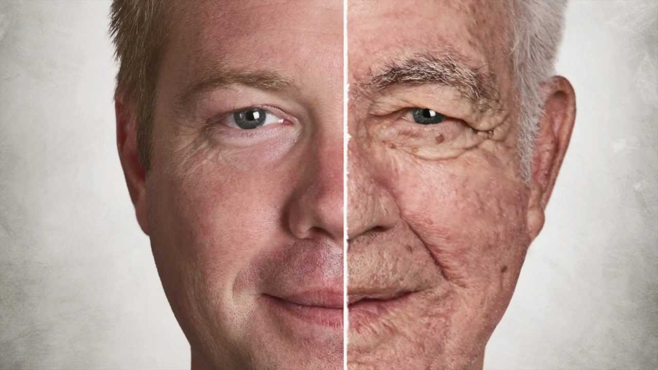
This week we will be referring back to our recent Scientist Spotlight on Linlin Sun to understand skin physiology through the viewpoint of aging.  Aging turns out to be a very helpful context for our studies, since all of our major skin cells and chemicals have distinct roles in the aging process.  As a result, we can get a fairly comprehensive overview of skin anatomy and physiology through this one subject - aging!  We will additionally be working more extensively with an academic paper in order to practice our scientific article reading skills.

Given all of the above, we will seek to...

  • Use evidence from a scientific article to describe the impacts of aging on skin cells, skin molecules, and normal skin physiology.

  • Demonstrate increased comfort and proficiency while reading from a scientific primary source.

A Note About Studying Aging & the Skin

Our society puts a lot of pressure on us - particularly those who identify as women - to prevent and hide certain signs of aging in our skin.  This can create a lot of stress for some individuals!  To be clear, aging, including aging of the skin, can be a wonderful and beautiful process, and not necessarily a process to fear or avoid.  In this module, we will simply be looking at some natural changes that occur in the skin during our lives.  Some of those changes we might wish to prevent, since they involve a decrease in some of skin's functions.  However, there is no presumption or implication that there's a biological basis for wishing to prevent these changes for cosmetic reasons!

skin physio-week6


Aging is still something of a mysterious process from an anatomy and physiology perspective, but it is a fascinating one to explore!  View the video below for an overview of what physiologists currently think about aging (or copy and paste this address: https://www.youtube.com/watch?v=GASaqPv0t0g)

Afterward, compose a brief reflection (approximately 4-5 sentences) on...

What were some of the main themes from the video in terms of physiological changes that happen during aging?

         After watching video, I realized that health and aging depend on what we eat especially contain ultra-processed foods in our daily life. The physiological changes that happen during aging caused by GM foods that we consume every day which interacts harmful effects to our body. 

What confusions or questions come up for you after this video?

            It came across into my mind that how can I prevent from aging.  Mitochondria produce ATP which play important role and cellular process. I am wondering how I can maintain mitochondria healthy and prevent from deterioration.

What do you already know about what happens to the skin in aging?

The sun's ultraviolet light can cause major damage to the skin related to aging.  Eating ultra-processed foods also another reason that leading speed up the aging of body cells. Consume lots of trans fats and can promote the chronic inflammation that hastens the breakdown the body cells lead to aging. Consuming GM foods that caused harmful effects on the human body including DNA damage result in mitochondrial dysfunction. If all cells in the body lost their mitochondria, the cells, tissues and eventually organs inability to operate because it cannot produce energy for cell.


1997 

112 years 

oldest person in earth

human are build 

90 years capacity 

age- growing up 

others growing old 

scientific definition of aging 

interaction with sunlight - The sun's ultraviolet light can cause major damage to the skin. The outer layer of the skin has cells that contain the pigment melanin. Melanin protects skin from the sun's ultraviolet rays. These can burn the skin and reduce its elasticity, leading to premature aging.

air ,

 How aging interaction with diet -Research shows that eating ultra-processed foods can speed up the aging of your cells.

 Much of this is due to ultra-processed foods often containing a high amount of hydrogenated oils, which are full of trans fats and can promote the chronic inflammation that hastens the breakdown (or aging) of your cells.

structure and function of the molecule of the cell. 

recently 9 physiological treats 

1 - body genetic damage -DNA damage can also result in mitochondrial dysfunction, impaired autophagy, metabolic changes, and the triggering of cellular senescence (small circles). These live but physiologically altered cells are predicted to be a more potent driver of aging and disease.

2 DNA replicate -DNA Replication Stress and Aging

Replication stress may lead to genomic instability, aging, and cancer. Genomic instability may increase during aging due to replication pausing and stalling. Generally, different forms of DNA damage and DNA breakage increase in pausing/stalling of replication. 

mitochondria produce ATP (adenosine triphosphate). Main energy for all cellular process. 

program cell death. 

mitochondria dead -Mitochondrial biogenesis declines with age due to alterations in mitochondrial dynamics and inhibition of mitophagy, an autophagy process that removes dysfunctional mitochondria.

 Age-dependent abnormalities in mitochondrial quality control further weaken and impair mitochondrial function.

epi- genetic

older people -Alzheimer's 

harmful genetic alteration -causes: The biggest threat caused by GM foods is that they can have harmful effects on the human body. 

It is believed that consumption of these genetically engineered foods can cause the development of diseases which are immune to antibiotics.


What are GMOs?

Genetically Modified Organisms (GMOs) are created through the process of genetic engineering: a technology that takes DNA from one organism and moves it into another, creating new varieties of plants and animals that wouldn’t be found in nature.

Organic is Non-GMO

Organic is non-GMO because the use of GMOs is prohibited in organic production. For example, organic farmers cannot plant GMO seeds, organic livestock cannot eat GMO feed, and organic food manufacturers cannot use GMO ingredients.

What GMO crops are in the United States?

Corn: Corn is the most commonly grown crop in the United States, and most of it is GMO. ...

Soybean: 

Cotton: ...

Potato: ...

Papaya: ...

Summer Squash: ...

Canola: ...

Alfalfa:


decline as we age. 

package with chromosome! 

telomer become too short - process that halt cycle. 

cancer proliferation 

cutting short to replicate 

resize - without limit to other cells. 

other changes revolved 

stop being able to do 

toxic nutrients 

intercellular communication 

aging

longer life we know it 

exercise 

medicine 

gene therapy

live longer we already do 

no tell


how can you prevent mitochondria dead? 

Clinical trials have shown the utility of using oral replacement supplements, such as l-carnitine, alpha-lipoic acid (α-lipoic acid [1,2-dithiolane-3-pentanoic acid]), coenzyme Q10 (CoQ10 [ubiquinone]), reduced nicotinamide adenine dinucleotide (NADH), membrane phospholipids, and other supplements.

10 Ways You Can Boost Mitochondria

Mitochondria sustain human life because they generate the energy that powers our cells. By supporting mitochondrial fitness, we are supporting the optimal functioning of pretty much every cell and system in the body.

Boosting mitochondria can help us live a long and healthy life.

  1.  Calorie Restriction,
  2.  exercise 
  3. mitochondria nutrients 
  4. sleep 
  5. relaxation techniques 
  6. Red and near-infrared light can penetrate the skin and act on mitochondria through stimulation of a molecule called cytochrome C oxidase. A number of benefits have been described for red/near-infrared light therapy, including, for example, accelerated wound healing and increased capacity to decrease muscle damage and improve muscle recovery following exercise.
  7. Supporting NAD-NAD+ (from nicotinamide adenine dinucleotide) is a molecule derived from vitamin B3 found in every single cell in the body. NAD+ has a key role in mitochondrial function: It is the main molecule responsible for the delivery of the electrons that are extracted from food to the electron transport chain for ATP production.
  8. cold exposure 

  9. heat exposure
  10. sun light: Without forgetting that excessive unprotected sunbathing can be very harmful, it’s important to remember that the right amount of sun is fundamental for our healthA well-known effect of sunlight is the production of vitamin D in our skin. It turns out that vitamin D is necessary for mitochondrial activity and that vitamin D supplementation in vitamin D-deficient adults improves mitochondrial oxidative capacity in muscles. Furthermore, animal studies have shown that vitamin D promotes mitochondrial biogenesis and increases the oxidative capacity of mitochondria in muscles and brown fat.

Cancer is a generic term for many diseases

 Cancer is a generic term for many diseases in which cells escape regulatory signals. Uncontrolled growth, invasion into adjacent tissues, and colonization of other organs, if not treated early enough, are its hallmarks. Health suffers when tumors “rob” blood supply from the “normal” organs.


 Many tumors are benign, meaning they do not metastasize nor cause disease. A tumor becomes malignant, or cancerous, when it breaches the confines of its tissue, promotes angiogenesis, attracts the growth of capillaries, and metastasizes to other organs.

 he specific names of cancers reflect the tissue of origin. Cancers derived from epithelial cells are referred to as carcinomas. Cancer in myeloid tissue or blood cells form myelomas. Leukemias are cancers of white blood cells, whereas sarcomas derive from connective tissue. Cells in tumors differ both in structure and function. Some cells, called cancer stem cells, appear to be a subtype of cell responsible for uncontrolled growth. Recent research shows that contrary to what was previously assumed, tumors are not disorganized masses of cells, but have their own structures.

 Cancer treatments vary depending on the disease’s type and stage. Traditional approaches, including surgery, radiation, chemotherapy, and hormonal therapy, aim to remove or kill rapidly dividing cancer cells, but these strategies have their limitations. Depending on a tumor’s location, for example, cancer surgeons may be unable to remove it. Radiation and chemotherapy are difficult, and it is often impossible to target only the cancer cells. The treatments inevitably destroy healthy tissue as well. To address this, researchers are working on pharmaceuticals that can target specific proteins implicated in cancer-associated molecular pathways.

 Cells may begin to function abnormally, which may lead to diseases associated with aging, including arthritis, memory issues, and some cancers.

cancer

 Cancer treatments vary depending on the disease’s type and stage. Traditional approaches, including surgery, radiation, chemotherapy, and hormonal therapy, aim to remove or kill rapidly dividing cancer cells, but these strategies have their limitations.Depending on a tumor’s location, for example, cancer surgeons may be unable to remove it. Radiation and chemotherapy are difficult, and it is often impossible to target only the cancer cells. The treatments inevitably destroy healthy tissue as well. To address this, researchers are working on pharmaceuticals that can target specific proteins implicated in cancer-associated molecular pathways.

Aging

The progressive impact of aging on the body varies considerably among individuals, but Studies indicate, however, that exercise and healthy lifestyle choices can slow down the deterioration of the body that comes with old age. 


 region of thechromosone - 

cell are less able to redevide regenerate

shorting it time 

divide - regenerate 

transport the o2 and remove 

abnormally - associate iwth aging 

arthritis 

cancer 

memory issue 

healthy lifestyle can slow down the aging 

cancer - 

uncontrolled growth

colonization of other organ 

mutation is permanant change of the DNA 

DNA is decode - generate abnormal cell 

enviroment , infectious 

mordification -prolifiration 

accumate - loose to form normal tissue 

many tumor -

tomor become maglinant -cancerous 

metastatis 

4.22 

cancer the tissue of o


can myloid -mylomas 

lukemia -wbc

sarcoma related to connective tissue 

uncontrolled growth 

tumor are not disorganized 

tumor 

surgery

radiation 

remove or rapidly devide cancer 

depending on the location of cancer 

radion and chemotherapy difficut 

destroyed the healthly cell as well 


Skin Wound Healing Process

Nervous tissue

 By the end of this section, you will be able to:


Identify the classes of cells that make up nervous tissue

Discuss how nervous tissue mediates perception and response

Nervous tissue is characterized as being excitable and capable of sending and receiving electrochemical signals that provide the body with information. Two main classes of cells make up nervous tissue: the neuron and neuroglia (Figure 4.19). Neurons propagate information via electrochemical impulses, called action potentials,which are biochemically linked to the release of chemical signals. Neuroglia play an essential role in supporting neurons and modulating their information propagation.

Neurons display distinctive morphology, well suited to their role as conducting cells, with three main parts. The cell body includes most of the cytoplasm, the organelles, and the nucleus. Dendrites branch off the cell body and appear as thin extensions. A long “tail,” the axon, extends from the neuron body and can be wrapped in an insulating layer known as myelin, which is formed by accessory cells. The synapse is the gap between nerve cells, or between a nerve cell and its target, for example, a muscle or a gland, across which the impulse is transmitted by chemical compounds 

smooth muscle

 Smooth muscle tissue contraction is responsible for involuntary movements in the internal organs. It forms the contractile component of the digestive, urinary, and reproductive systems as well as the     airways and arteries. Each cell is spindle shaped with a single nucleus and no visible striations (Figure 4.18).

cardiac muscle

 Cardiac muscle forms the contractile walls of the heart. 

The cells of cardiac muscle, known as cardiomyocytes, also appear striated under the microscope. Unlike skeletal muscle fibers, cardiomyocytes are single cells typically with a single centrally located nucleus.  

 A principal characteristic of cardiomyocytes is that they contract on their own intrinsic rhythms without any external stimulation. Cardiomyocyte attach to one another with specialized cell junctions called intercalated discs. Intercalated discs have both anchoring junctions and gap junctions. Attached cells form long, branching cardiac muscle fibers that are, essentially, a mechanical and electrochemical syncytium allowing the cells to synchronize their actions. The cardiac muscle pumps blood through the body and is under involuntary control. The attachment junctions hold adjacent cells together across the dynamic pressures changes of the cardiac cycle.

skeletal Muscle tissue

 Muscle tissue is classified into three types according to structure and function: skeletal, cardiac, and smooth 

skeletal :Long cylindrical fiber, striated, many peripherally located nuclei Voluntary movement, produces heat, protects organsLocation=> Attached to bones and around entrance points to body  (e.g., mouth, anus)

Cardiac muscle Short, branched, striated, single central nucleus Contracts to pump blood- location=> Heart


Smooth Short, spindle-shaped, no evident striation, single nucleus in each fiber Involuntary movement, moves food, involuntary control of respiration, moves secretions, regulates flow of blood in arteries by contraction-location : Walls of major organs and passageways 


Skeletal muscle is attached to bones and its contraction makes possible locomotion, facial expressions, posture, and other voluntary movements of the body.

Forty percent of your body mass is made up of skeletal muscle. Skeletal muscles generate heat as a byproduct of their contraction and thus participate in thermal homeostasis.

Shivering is an involuntary contraction of skeletal muscles in response to perceived lower than normal body temperature. The muscle cell, or myocyte, develops from myoblasts derived from the mesoderm. Myocytes and their numbers remain relatively constant throughout life. Skeletal muscle tissue is arranged in bundles surrounded by connective tissue.

Under the light microscope, muscle cells appear striated with many nuclei squeezed along the membranes. The striation is due to the regular alternation of the contractile proteins actin and myosin, along with the structural proteins that couple the contractile proteins to connective tissues.The cells are multinucleated as a result of the fusion of the many myoblasts that fuse to form each long muscle fiber.

Wound healing versus fibrosis

 U